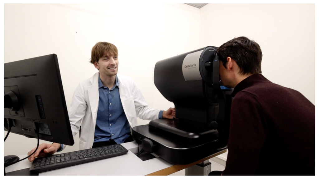
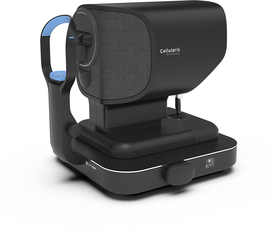
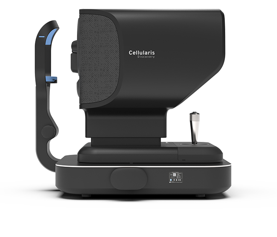
See the invisible

EarlySight aims to reduce vision loss by enabling precise detection and better monitoring of eye diseases. The company develops high-performance eye imaging instruments empowering doctors to see disease impacts that are invisible with the standard of care.

Reducing vision loss

Blindness and severe vision impairments consecutive to eye disorders affect more than 200 million people worldwide. Indeed, current diagnoses are made too late, and treatments are not optimal for many diseases. The main reason is the difficulty of accurately detecting and monitoring the disease as well as assessing the treatment effect. This is due to the limitations of the standard imaging instruments unable to visualize the eye tissues with sufficient details.

At EarlySight, we have developed a unique ophthalmic camera that can observe the retina in detail, as if under a microscope. This advanced optical instrument creates images of the individual cells impacted by diseases. An AI software is in development to analyze these images and detect and monitor disease biomarkers. The perspective to precisely measure the degenerative process opens new avenues in the diagnosis, monitoring and treatment of disorders affecting the back of the eye.

Standard SLO instrument_RPEsmallEarlySight technology_RPEsmall

Cellular resolution

Look at every detail in the eye

Digital data

AI-based objective structural measurement

Explore disease inception

Unique access to the deep eye layer (RPE)

Cellularis Discovery

The disruptive ophthalmic camera from EarlySight, delivers cellular level images of various structures of the retina and optic disc to explore and capture the eye anatomy in a completely new perspective.

See the invisible

Disruptive infrared transscleral flood illumination reveals hidden eye structures such as RPE.

Outstanding resolution

With adaptive optics, look at single cells composing the tissue, as if under a microscope. Observe the retina anatomy in detail.

Targeted imaging modes

Easy switch between retinal pigment epithelium, photoreceptors, nerve fiber layer or retinal vessels image

Fundus overview

Obtain widefield fundus overview image simultaneously with the cellular level image to easily locate the imaged area within the eye.

Easy-to-use

Manual and smooth operation with joystick allow for quick and efficient examinations.

Follow-up

Automatic storage of image location within the eye to easily perform follow-up examinations.

Testimonials

EarlySight’s Cellularis platform has already been endorsed by several renowned eye doctors.
Prof. Dr. med. Dr. phil. Michael A. Thiel
Chefarzt Augenklinik, Bereichsleiter (COO) Spezialkliniken und Institute LUKS Gruppe, Switzerland
Read More
The Cellularis platform offers an entirely new approach to visualizing the retinal pigment epithelial cells. This technique will offer exciting new options to understand retinal diseases.
Prof. Dr. med. Dr. phil. Francine Behar-Cohen
Head of the Department of Ophthalmology of Hôtel-Dieu – University of Paris Descartes Hospital, France.
Read More
It allows to image a cell layer that has never been directly visualized. As the RPE is the priamary site of almost all retinal diseases, we can expect that it will become a must in our daily practice.
Prof. Dr. Jeffrey M. Liebmann
Glaucoma Service Director, and Vice-Chair for the Department of Ophthalmology at Columbia University Medical Center, US.
Read More
Cellularis imaging has the potential to facilitate early diagnosis of retinal disorders affecting photoreceptors and the retinal pigment epithelium, monitor disease progression, and assess the outcomes of novel therapeutic interventions.
Prof. Dr.-Ing. Dr. med Sebastian Wolf
Chefarzt und Co-Klinikdirektor at University Department of Ophthalmology, Inselspital, University of Bern, Switzerland
Read More
Cellularis imaging has the potential to address critical unmet medical needs in retinal disease such as early diagnosis, disease progression, therapy monitoring, and risk profiling. I am impressed by the ease of use and acquisition speed of the device to obtain high-resolution images.
Prof. Dr. Jeffrey M. Liebmann
Glaucoma Service Director, and Vice-Chair for the Department of Ophthalmology at Columbia University Medical Center, US.
Read More
In the field of glaucoma, Cellularis imaging could provide extremely high-resolution imaging of the retinal nerve fiber layer, which has implications for both glaucoma clinical trials and earlier detection of disease progression.
Prof. Dr. med. Dr. phil. Francine Behar-Cohen
Head of the Department of Ophthalmology of Hôtel-Dieu – University of Paris Descartes Hospital, France.
Read More
In my experience in patients with CSCR, it allows to see abnormalities in the RPE that other imaging modalities cannot detect. We know need to evaluate whether these changes have prognostic values.
Previous
Next

Supporters

We are deeply appreciative of the support and trust that our partners have shown us.

Latest news